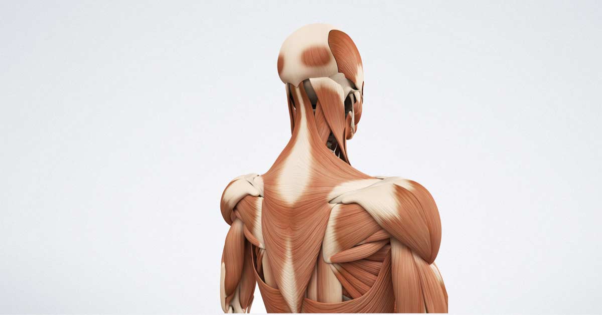
Back Of Neck Anatomy Muscles - Zyfsa4xa1qbmkm - Muscles of the shoulder and back laminated anatomy chart.
byAdrienne Black•
0
Back Of Neck Anatomy Muscles - Zyfsa4xa1qbmkm - Muscles of the shoulder and back laminated anatomy chart.. This article gives an overview of the back's structure and its major muscles. Anterior muscles of the neck. Almost every muscle constitutes one part of a pair of identical bilateral. Back muscles are arranged in several layers, so they are divided into deep and superficial, which, in turn, are arranged in two layers. The superficial group acts on upper limbs and.
The neck has no external bone protective structures, so it is quite mobile. The anterior muscles of the neck facilitate swallowing and speech. The superficial group acts on upper limbs and. The head rests on the top part of the vertebral column, with the skull joining at c1. As you know, the neck is the part of the body that sits between the head and torso.
Yuurdxq9iwdjlm from simplifaster.com Intermediate back muscles and c. Digastric, mylohyoid, geniohyoid, stylohyoid infrahyoid muscles: They are divided into three groups, as shown below. Muscles make up a large part of the anatomy (structure) of the back. Neck mobility is necessary primarily to rotate the head and keep the head upright. Human muscle system, the muscles of the human body that work the skeletal system, that are under voluntary control, and that the following sections provide a basic framework for the understanding of gross human muscular anatomy, with descriptions of the large muscle groups and their actions. Integrates anatomy and physiology of cells, tissues, organs, the systems of the human body, and mechanisms responsible for homeostasis. The neck muscles, including the sternocleidomastoid and the trapezius, are responsible for the gross motor movement in the muscular system of the head and neck.
The posterior muscles of the neck are primarily concerned with head movements, like extension. Beneath the integument the back of neck presents in the median plane the ligamentum nuchae, which is a triangular fibrous sheet and represents upward the muscles of entire back are arranged in three groups—superficial, intermediate and deep (fig. Together they extend neck, and individually they draw and rotate head to one side i.e. The muscles of the shoulder and back chart shows how the many layers of muscle in the shoulder and back are intertwined with the other relevant systems and muscles in adjacent areas like the spine and neck. They move the head in every direction, pulling the skull and jaw towards the shoulders, spine, and scapula. Muscles make up a large part of the anatomy (structure) of the back. Sternohyoid, sternothyroid, thyrohyoid, omohyoid anterior vertebral muscles: There are many muscles around the neck that help to support the cervical spine and allow you to move your head in different directions. Intermediate layer of back muscles. This article describes the anatomy of the head and neck of the human body, including the brain, bones, muscles, blood vessels, nerves, glands, nose, mouth, teeth, tongue, and throat. Watch cervical muscle anatomy animation. The suprahyoid muscles originate from above the hyoid bone in the chin region. 12 photos of the anatomy of neck muscles.
Week 2 anatomy (back/neck muscles). Muscles make up a large part of the anatomy (structure) of the back. There are around 650 skeletal muscles within the typical human body. Intermediate layer of back muscles. The neck muscles, including the sternocleidomastoid and the trapezius, are responsible for the gross motor movement in the muscular system of the head and neck.
Hubspot from The anterior and middle scalenes originate from the transverse processes of certain cervical vertebrae and attach to the first rib. Back pain is common and might be caused by a problem with a muscle. We will attempt to provide a simplified overview of this complex anatomy. As you know, the neck is the part of the body that sits between the head and torso. Human anatomy for muscle, reproductive, and skeleton. Only two of the more obvious and superficial neck muscles are. Last update october 2, 2020. The anterior muscles of the neck facilitate swallowing and speech.
The suprahyoid muscles originate from above the hyoid bone in the chin region.
Tutorials and quizzes on the anatomy and actions of the back muscles (iliocostalis, longissimus, spinalis, multifidus, and quadratus lumborum), using interactive animations, diagrams, and illustrations. Intermediate back muscles and c. This is a table of skeletal muscles of the human anatomy. Neck muscles help support the cervical spine and contribute to movements of the head, neck, upper back, and posterior longitudinal ligament (pll). The posterior muscles of the neck are primarily concerned with head movements, like extension. The anterior muscles of the neck facilitate swallowing and speech. Here the extrinsic back muscles are classified into logical subgroups to facilitate knowledge. The suprahyoid muscles originate from above the hyoid bone in the chin region. Last update october 2, 2020. Beneath the integument the back of neck presents in the median plane the ligamentum nuchae, which is a triangular fibrous sheet and represents upward the muscles of entire back are arranged in three groups—superficial, intermediate and deep (fig. William is a final year medical student in australia who has taught anatomy to tertiary science and medical students since 2010. The superficial group acts on upper limbs and. This article gives an overview of the back's structure and its major muscles.
This article describes the anatomy of the head and neck of the human body, including the brain, bones, muscles, blood vessels, nerves, glands, nose, mouth, teeth, tongue, and throat. Digastric, mylohyoid, geniohyoid, stylohyoid infrahyoid muscles: Last update october 2, 2020. Watch cervical muscle anatomy animation. The suprahyoid muscles originate from above the hyoid bone in the chin region.
3skppra4yhoplm from media.sciencephoto.com We will attempt to provide a simplified overview of this complex anatomy. Tutorials and quizzes on the anatomy and actions of the back muscles (iliocostalis, longissimus, spinalis, multifidus, and quadratus lumborum), using interactive animations, diagrams, and illustrations. The anterior and middle scalenes originate from the transverse processes of certain cervical vertebrae and attach to the first rib. The back anatomy includes the latissimus dorsi, trapezius, erector spinae, rhomboid, and the teres major. Some neck muscles attach to the clavicles. It's buried under the sternomastoid anteriorly and by. As you know, the neck is the part of the body that sits between the head and torso. The anterior muscles of the neck facilitate swallowing and speech.
Alle muscles are detailed described incl.
They start at the top of the neck and go down to the tailbone. Bones of the neck picture. The pll starts at c2 and goes down the back of the vertebral bodies and intervertebral discs. The anterior and middle scalenes originate from the transverse processes of certain cervical vertebrae and attach to the first rib. The superficial group acts on upper limbs and. Here the extrinsic back muscles are classified into logical subgroups to facilitate knowledge. The anterior muscles of the neck facilitate swallowing and speech. There are around 650 skeletal muscles within the typical human body. Cervical spine anatomy is quite complex. The suprahyoid muscles originate from above the hyoid bone in the chin region. Human muscle system, the muscles of the human body that work the skeletal system, that are under voluntary control, and that the following sections provide a basic framework for the understanding of gross human muscular anatomy, with descriptions of the large muscle groups and their actions. The head rests on the top part of the vertebral column, with the skull joining at c1. Several other muscles of the back also extend up to the neck region and are partly connected with the cervical part of the vertebral column, including the trapezius, levator scapulae, splenius, iliocostalis, longissimus, rotatores, semispinalis, interspinales, and intertransversarii muscles.
This article gives an overview of the back's structure and its major muscles back of neck anatomy. William is a final year medical student in australia who has taught anatomy to tertiary science and medical students since 2010.